
Pathology
Conditions in and around the hip
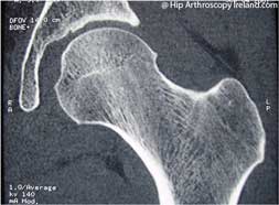
- Routine investigations for patients with suspected hip pathology include:
- AP pelvis x-ray and a true lateral view of the femoral neck
- High resolution 3-D reconstructed CT scan of the hip
- MRI of the hip with saggital, coronal and axial reconstructions
- MR arthrogram of the hip with saggital, coronal and axial reconstructions
- AP pelvis x-ray and a true lateral view of the femoral neck
 |
|
 |
|
 |
|
 |
 |
|
 |
 |
 |
 |
|
 |
|
Acetabular labrum
- The labrum is a ring of fibrocartilage ‘grisel’ around the rim of the socket. It is frequently torn by injury or as part of femoroacetabular impingement (FAI). It may also degenerate with wear and tear
 |
|
 |
Ligamentum Teres
- The ligamentum teres is a structure deep inside the hip joining the femoral head to the base of the socket
- Acute injuries may cause ligamentum teres tears or they can gradually occur with wear and tear
 |
|
Articular cartilage
- Articular cartilage is the slippery surface of the joint. The smooth surfaces of the ball and socket which glide over one another as the joint moves.
 |
|
 |
|
 |
|
 |
|
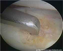
Acetabular Microfracture
- One of the treatments for localised full thickness cartilage loss in an area is acetabular microfracture
- This causes bleeding into the defect which stimulates a healing response. A fibrocartilage scar forms over the area
 |
Osteoarthritis
- While short term relief of mechanical symptoms can often be gained, hip arthroscopy is not a long-term solution to arthritis
 |
|
 |
|
 |
|
 |
|
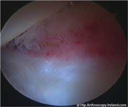
Synovitis
 |
|
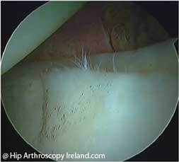
Femoroacetabular impingement lesions
- These ‘bumps’ on the femoral neck impact on the rim of the socket causing pain, labral tears and cartilage damage
- They are painless, but cause symptoms by the damage they inflict
- Excision of the impingement lesion is part of the treatment of femoroacetabular impingement, along with treatment of the damaged labrum and cartilage
- These are some impingement lesions at the head/neck junction viewed at hip arthroscopy prior to removal
 |
 |
 |
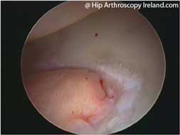
Loose bodies
- Loose bodies frequently cause painful mechanical symptoms inside the hip
- They may arise from a loose piece of bone after an injury, or as part of an underlying condition such as arthritis or synovial chondromatosis
- Loose bodies viewed at hip arthroscopy and removal
 |
 |
 |